

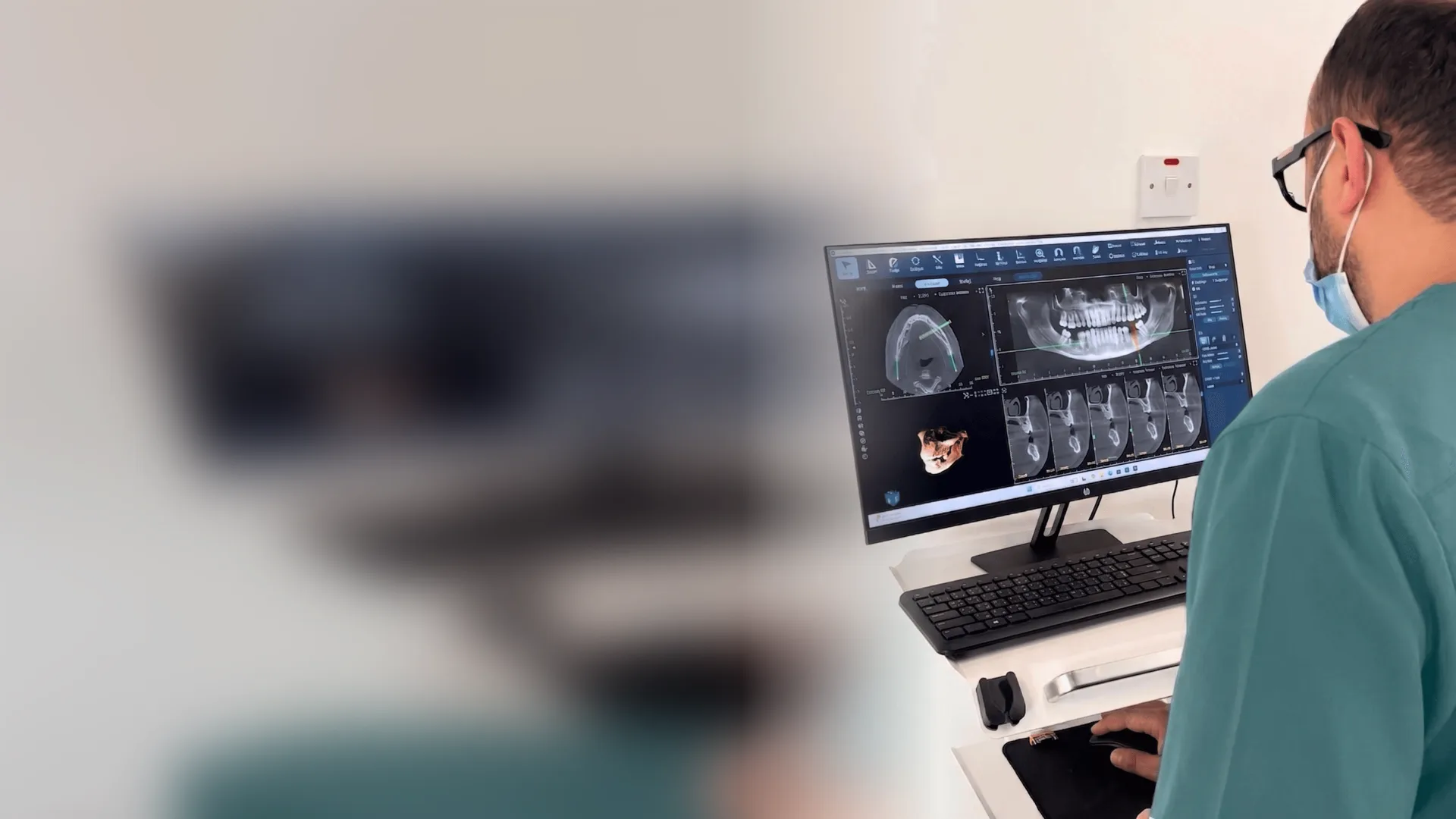
A CBCT dental scan (cone beam computed tomography) is a 3D X-ray of your teeth and jaws taken in a single rotation that lasts well under a minute. Unlike a normal 2D X-ray, it shows bone width, nerve position, root shape and hidden problems from every angle. It is the standard tool for planning dental implants and for assessing impacted wisdom teeth, complex root canal cases, cysts and some orthodontic problems. It is not a routine scan: a good dentist orders it only when the 3D information will change the treatment, and chooses the smallest field of view that answers the question.
People usually search for 3D dental imaging after a dentist mentions it: before an implant, before removing a wisdom tooth, or when a toothache doesn't match what the normal X-ray shows. The questions are always similar. What exactly is a CBCT dental scan? Is the radiation safe? Do I really need it, or is it an extra?
Those are fair questions, and the answers are clearer than most people expect. Here is what a CBCT scan shows, when it genuinely helps, when it doesn't, and what happens on the day.
What is a CBCT dental scan?
CBCT stands for cone beam computed tomography. The machine sends a cone-shaped X-ray beam through the area being examined while it rotates once around your head. In that single turn it captures hundreds of small images, and software rebuilds them into a 3D volume.
On the screen, the dentist can then slice through your jaw in any direction, measure distances to a fraction of a millimetre, and rotate the model to look at a tooth from the side, from above or from inside the bone. A dental CBCT, as the US FDA explains, is designed specifically for the teeth and jaws, which is why it is quicker and usually uses far less radiation than a hospital CT scan of the same region.
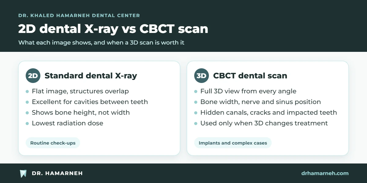
2D X-ray vs 3D dental imaging
Traditional dental X-rays, like the small bitewing films and the wide panoramic image, are still excellent for many jobs: finding cavities between teeth, checking bone levels, and getting an overview of the mouth. Their limitation is that they compress a three-dimensional jaw into a flat picture. Structures overlap, and depth is lost.
| Question | 2D X-ray | CBCT dental scan |
|---|---|---|
| Cavities between back teeth | Very good (bitewings) | Not the right tool |
| Bone height | Yes | Yes |
| Bone width and shape | No | Yes |
| Exact position of the nerve in the lower jaw | Approximate | Precise, in 3D |
| Distance to the sinus floor | Approximate | Precise |
| Extra or curved roots and hidden canals | Often missed | Usually visible |
| Impacted teeth relative to neighbours | Overlapping | Clear from every angle |
| Radiation | Lowest | Higher than 2D, much lower than medical CT in most cases |
So CBCT does not replace normal X-rays. It answers the questions they can't.
When you need a CBCT dental scan
Dental implant planning
This is the most common reason. Before placing an implant, the dentist needs to know how much bone there is, how wide and how dense it is, and exactly where the nerve and sinus are. A CBCT scan lets us choose the implant size and angle on the computer first, and in suitable cases design a surgical guide that transfers the plan to the mouth. That is the basis of our precision implant dentistry.
Impacted wisdom teeth and canines
A lower wisdom tooth can sit very close to the nerve that supplies feeling to the lip and chin. When a panoramic X-ray suggests that risk, 3D dental imaging shows the real relationship and helps plan a safer removal. Impacted upper canines, which are common in teenagers, are located the same way before orthodontic treatment brings them into place.
Difficult root canal cases
Some teeth have extra or curved canals, cracks, or infection that doesn't show on a 2D film. When a tooth keeps hurting after treatment, or symptoms don't make sense, a small-field CBCT can reveal the cause.
Cysts, lesions and jaw problems
Shadows seen on a normal X-ray can be measured and located precisely in 3D, which helps decide whether they need monitoring or treatment, and whether a specialist referral is needed.
Selected orthodontic and jaw joint cases
CBCT is not used for every orthodontic patient, but it helps in complex cases: impacted teeth, root resorption, narrow bone, or certain jaw joint (TMJ) and airway assessments.
When you don't need a CBCT scan
A responsible clinic does not scan everyone. You usually do not need 3D imaging for:
- A routine check-up or cleaning.
- Checking for ordinary cavities, where bitewing X-rays are better.
- Simple fillings, most extractions and straightforward root canals.
- Mild orthodontic cases with no impacted teeth or unusual findings.
This follows a principle that international guidelines, including the American Dental Association guidance on dental imaging, share: every X-ray must be justified by a question that will change your care. If a dentist suggests a CBCT dental scan, it is fair to ask what they expect to find and how it will affect the plan.
Radiation from a CBCT scan: the honest numbers
Radiation is measured in microsieverts (µSv). Doses vary a lot between machines and settings, which is why ranges are more honest than single numbers. According to the European SEDENTEXCT project on CBCT radiation doses:
- A panoramic X-ray is typically in the range of a few to about 25 µSv.
- A small-field dental CBCT, covering a few teeth or one jaw, is often in the tens of µSv, though some machines and settings give more.
- A large-field CBCT covering both jaws and more of the face can reach a few hundred µSv.
- For comparison, everyone receives natural background radiation from the environment, on average around 2,400 µSv a year worldwide, or roughly 6 to 7 µSv every day.
Two things keep the dose as low as possible. First, choosing the smallest field of view that answers the question: scanning only the area of a planned implant, not the whole skull. Second, using settings matched to the task, rather than maximum resolution every time. Children and teenagers are more sensitive to radiation, so for them the justification needs to be even stronger.
If you are pregnant or might be, tell the dentist before any X-ray. Most non-urgent imaging can wait.
Book an assessment in Khalda. We will tell you whether 3D imaging is needed in your case, and why.
What happens during a CBCT dental scan
- Preparation. You remove earrings, glasses, hairpins, removable dentures and anything metal near the head and neck.
- Positioning. Depending on the machine, you stand or sit, rest your chin on a support and bite gently on a small guide. A protective collar may be used when it does not block the area being scanned.
- The scan. The arm rotates around your head once. The actual exposure usually takes between a few seconds and less than a minute. You just need to stay still.
- Results. The 3D images are ready within minutes. The dentist reviews them on screen and, ideally, shows you the findings.
There are no injections, no tunnel and no noise beyond the gentle hum of the machine. People who feel claustrophobic in hospital scanners are usually comfortable, because the machine is open.
From CBCT scan to treatment plan
A scan is only as useful as the person reading it. For an implant, the dentist uses the 3D data to mark the nerve, measure the bone, and place a virtual implant in the ideal position for the future crown. If bone is missing, the scan shows how much and where, so grafting can be planned in advance rather than discovered during surgery.
For a painful tooth, the dentist scrolls through thin slices to look for an extra canal, a crack or infection at the root tip. For an impacted tooth, the scan shows whether it touches the roots of neighbouring teeth and how best to approach it. In each case, you should leave knowing what was found and what it means for your options.
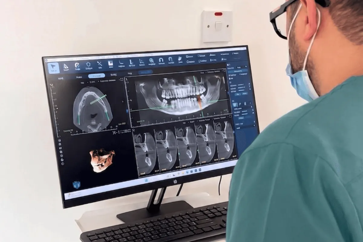
3D dental imaging at our clinic in Khalda
At Dr. Khaled Hamarneh Dental Center, 3D imaging is part of how we plan implants and complex cases, not a routine add-on. We use it when the answer will change your treatment, we choose the smallest area that answers the question, and we walk you through the images on screen so you understand your own case.
If you already have a CBCT scan from another centre, bring it on a USB or share the file. If it is recent and covers the right area, there may be no need to repeat it. You can read more about our team, or see how we approach urgent dental problems.
Minas Towers, 3rd Floor, Medical City Street, Khalda. Saturday to Thursday, 10:30 am to 7:00 pm.
Questions patients ask us
What is a CBCT dental scan?
A CBCT dental scan is a 3D X-ray of the teeth and jaws, taken by a machine that rotates once around the head. Software turns the images into a 3D model that shows bone, nerves, roots and hidden problems from every angle.
Is a CBCT scan the same as a CT scan?
They are related but not the same. A dental CBCT uses a cone-shaped beam designed for the teeth and jaws, is faster, and in most cases uses considerably less radiation than a hospital CT scan of the same area.
Is the radiation from a CBCT dental scan safe?
When it is justified and the smallest suitable field of view is used, the dose is low. A small-field dental CBCT is often in the range of tens of microsieverts, which is comparable to a few days to a few weeks of natural background radiation, though doses vary by machine and settings.
Do I need a CBCT scan before a dental implant?
In most cases, yes. A CBCT scan shows the width and quality of the bone and the exact position of the nerve and sinus, which allows the implant to be planned precisely and safely.
How long does a CBCT dental scan take?
The scan itself takes from a few seconds to under a minute. Including preparation and positioning, the whole visit for the scan usually takes about 10 to 15 minutes.
Can I have a CBCT scan if I am pregnant?
Tell your dentist if you are or might be pregnant. Non-urgent imaging is usually postponed. If imaging is essential, the dentist will weigh the need carefully and keep the dose to a minimum.
Is a CBCT scan suitable for children?
Only when there is a clear reason, such as locating an impacted tooth or assessing an injury. Children are more sensitive to radiation, so the justification must be strong and the smallest possible field of view is used.
Where can I get 3D dental imaging in Khalda, Amman?
Dr. Khaled Hamarneh Dental Center in Khalda uses 3D dental imaging to plan implants and complex cases when it is clinically needed. Book an assessment online, call or WhatsApp 0791818796.